ilustrações, clipart, desenhos animados e ícones de tecido ósseo compacto humano, estrutura do tecido ósseo ou tecido ósseo, osteon - histologia
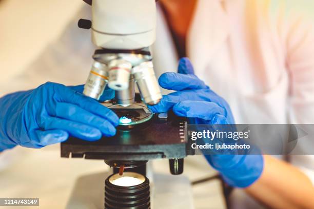
microscópio moderno com sistema de imagem digital em laboratório - histologia - fotografias e filmes do acervo
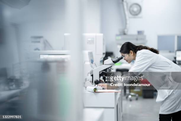
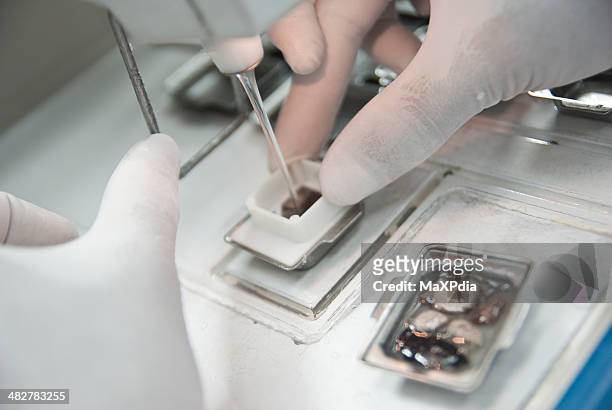
mulheres cientistas pesquisam e procuram microscópio em um laboratório - histologia - fotografias e filmes do acervo
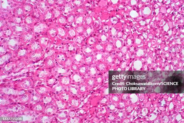
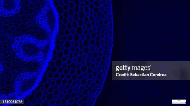
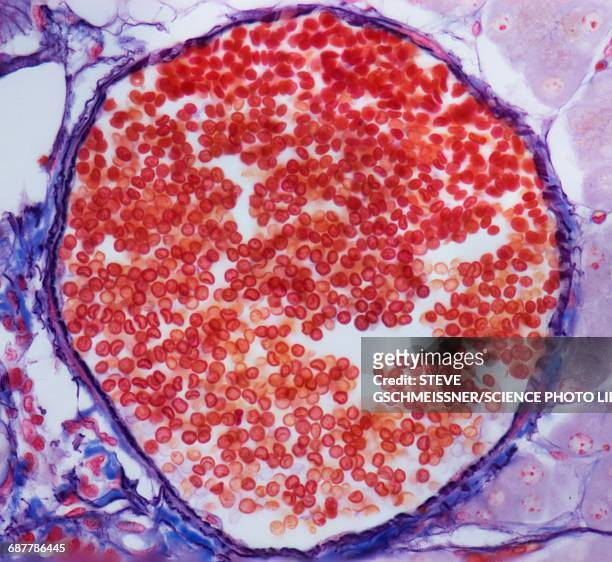
immunofluorescent photomicrograph, organs samples, histological examination, histopathology on the microscope - histologia - fotografias e filmes do acervo
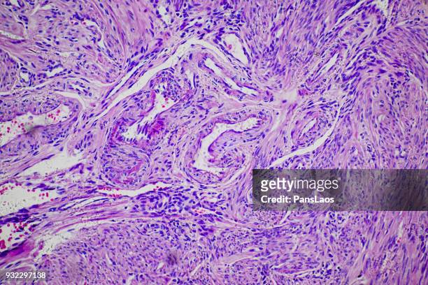
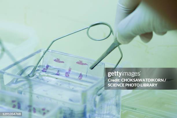
slide demonstrating breast tissue with ductal carcinoma. histopathology on the microscope. immunofluorescent photomicrograph, organs samples, histological examination, - histologia - fotografias e filmes do acervo
microscopic photo of a professionally prepared slide demonstrating breast tissue with ductal carcinoma. - histologia - fotografias e filmes do acervo
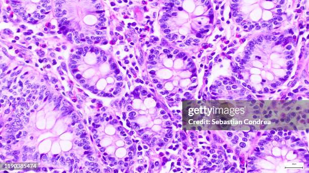
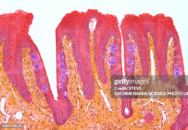
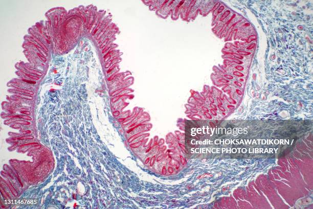
large intestine--mucosa or lining with very abundant goblet cells, 100x - histologia - fotografias e filmes do acervo
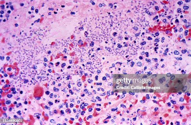
microscope of adenoid cystic carcinoma, rare type of cancer exist in many different body sites. this tumor occurs in the salivary glands, - histologia - fotografias e filmes do acervo
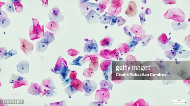
squamous epithelial cells of human cervix under the microscope view. pap smear test is a procedure to test for cervical cancer in women - histologia - fotografias e filmes do acervo
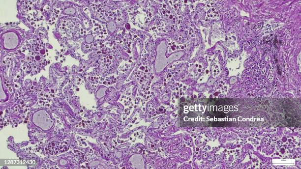
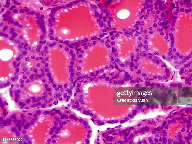
pancreas. serous acini, immunofluorescent photomicrograph, organs samples, histological examination, histopathology on the microscope. - histologia - fotografias e filmes do acervo
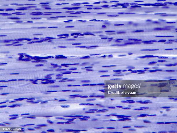
microscopy photography. cardiac muscle section, immunofluorescent photomicrograph, organs samples, histological examination, histopathology on the microscope. - histologia - fotografias e filmes do acervo
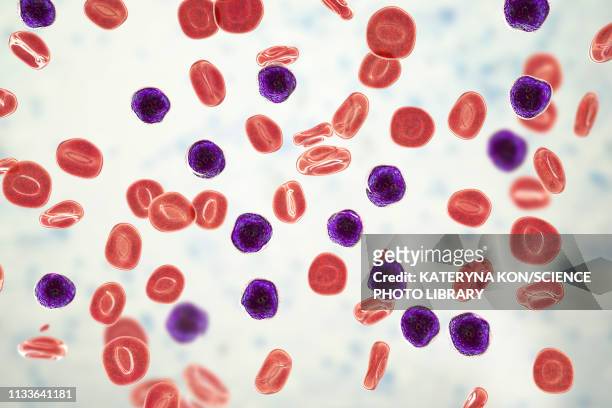
ilustrações, clipart, desenhos animados e ícones de acute lymphoblastic leukaemia, illustration - histologia
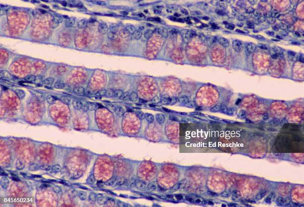
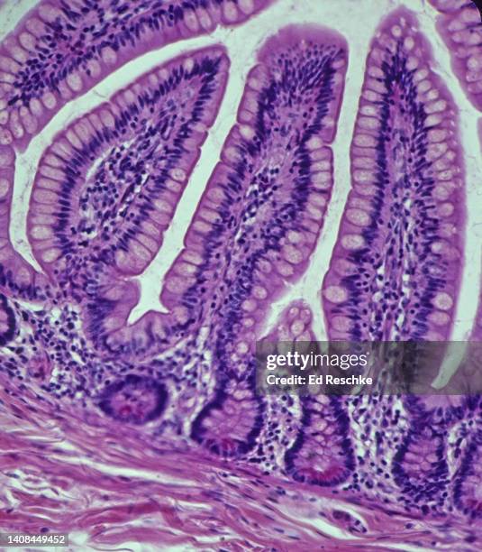
villi in the small intestine (ileum, human), simple columnar epithelium and intestinal glands, 50x - histologia - fotografias e filmes do acervo
small foramen magnum meningioma seen after gadolinium injection on axial t1 mri image - histologia - fotografias e filmes do acervo
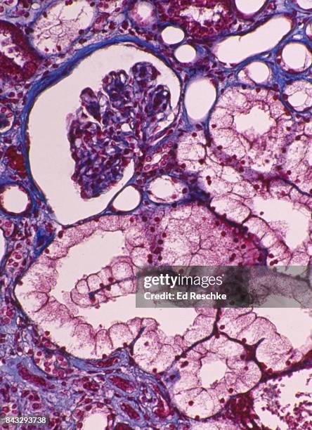
histological evaluation of the developing trabecular bone. goldner's trichrome staining with the blue/green colour representing mineralized tissue. immunofluorescent photomicrograph, organs samples. - histologia - fotografias e filmes do acervo
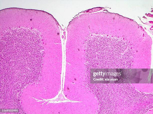
cross section of the cerebellum and nerve human under the microscope for education in lab - histologia - fotografias e filmes do acervo
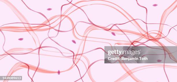
skeletal muscle fibres longitudinal section,40x light micrograph - histologia - fotografias e filmes do acervo
ilustrações, clipart, desenhos animados e ícones de pyramidal neurons in the cerebral cortex, illustration - histologia
histology (or microscopic anatomy) lining a bronchus, pseudostratified ciliated columnar epithelium, 100x - histologia - fotografias e filmes do acervo
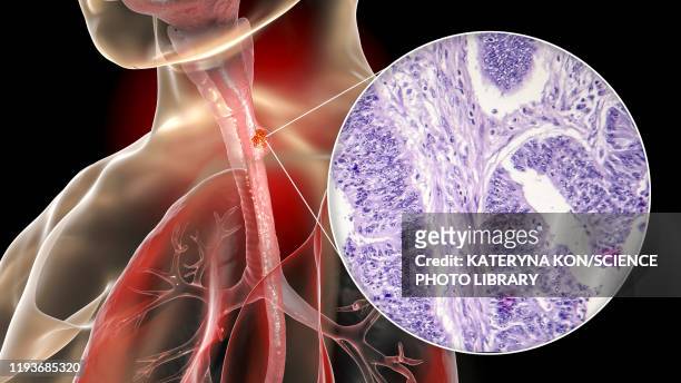
ilustrações, clipart, desenhos animados e ícones de oesophageal cancer, illustration and micrograph - histologia
ilustrações, clipart, desenhos animados e ícones de oesophageal cancer, illustration and micrograph - histologia